|
The Colour Group conducts research in such diverse areas as colour reproduction, computer vision, colour measurement, image enhancement, image retreival, and device characterisation/calibration. The unifying theme of all these research areas is colour. Thus to understand any of our research it is helpful to have a basic understanding of what colour is and how it is perceived both by ourselves, and by electronic devices such as colour cameras and colour scanners.
Our sensation of colour is a result of the processing of light energy first by the eye and later by the brain. The exact nature of this processing is complex, and indeed not yet fully understood. Fortunately, to gain an understanding of many of the problems we study, a very simple model of image formation suffices. Figure 1 is an illustration of a simple model of image formation. Light energy is emitted from a source (for example, the sun, or a tungsten filament light bulb) and is then incident upon a surface. Some, or all of the energy in the incident light is reflected from this surface and enters the eye. At the eye light is focused by a lens onto a membrane at the back of the eye, called the retina. On the surface of the retina are many light sensitive cells (called cones) which emit a response when light is incident upon them. There are three types of cone cells differentiated by how they respond to light energy. It is the responses of these three different cell types to light energy which are the basis of our colour perception.
|
|
| (1) |
The function ![]() is called the colour signal and it is this signal
which is focused by the lens at the retina, and to which
the light sensitive cone cells respond. The response of a cone cell depends
on how much energy is present in the light incident upon it and
importantly a cone cell can respond more or less to a given quantum of light
energy depending on the wavelength at which that
quantum is emitted. That is, the response of the cone cells to light energy
is a function of the wavelength at which the energy is emitted.
The three types of cone cells differ in how they respond to light energy at
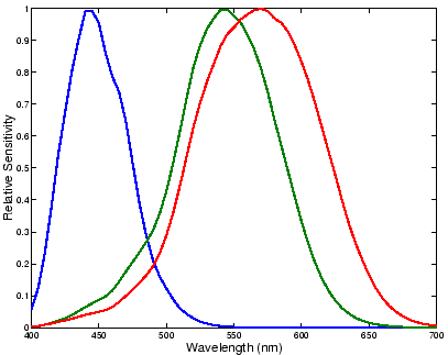
different wavelengths. Figure 4 shows the relative response of the
three different cone cells as a function of wavelength. The cone cells are
often called long-, medium-, or short-, wavelength sensitive cells
since to a first approximation, they are preferrentially sensitive to light
in one of these three wavelength regions of the visible spectrum.
is called the colour signal and it is this signal
which is focused by the lens at the retina, and to which
the light sensitive cone cells respond. The response of a cone cell depends
on how much energy is present in the light incident upon it and
importantly a cone cell can respond more or less to a given quantum of light
energy depending on the wavelength at which that
quantum is emitted. That is, the response of the cone cells to light energy
is a function of the wavelength at which the energy is emitted.
The three types of cone cells differ in how they respond to light energy at
different wavelengths. Figure 4 shows the relative response of the
three different cone cells as a function of wavelength. The cone cells are
often called long-, medium-, or short-, wavelength sensitive cells
since to a first approximation, they are preferrentially sensitive to light
in one of these three wavelength regions of the visible spectrum.
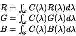
The total response of the cone cells are the sum of their response to light
energy at all wavelengths to which they are sensitive. Mathematically we can
write these sums as integrals and if we denote the response of the three types
of cone cell as ![]() ,
, ![]() , and
, and ![]() we have:
we have:
 |
(2) |
where ![]() ,
, ![]() , and
, and ![]() represent the relative
sensitivities of the three different cone cells and
represent the relative
sensitivities of the three different cone cells and ![]() is the wavelength range for which the cells have non-zero response. It is this
triplet of cone cell responses which is the basis for all subsequent
visual processing by the eye and the brain which result eventually in our
sensation of colour.
is the wavelength range for which the cells have non-zero response. It is this
triplet of cone cell responses which is the basis for all subsequent
visual processing by the eye and the brain which result eventually in our
sensation of colour.
 |
(3) |
Equations (2) and (3) represent a mathematical description of the image formation process at the eye and in colour imaging devices. In the case of our own visual system however, there is a great deal of subsequent processing of the initial cone responses, processing which leads eventually to our perception of colour and more generally to our visual perception as a whole. Notwithstanding this, this simple model of image formation forms the starting point for much of the research which is undertaken in the group, some of which is described in more detail in these pages.